CT and colorful 3D scans combine to create a stunningly detailed depiction of a mummy of a girl. She was only about five years old when she passed away 2,000 years ago. Her body was wrapped in fine linen and she was laid to rest with a Roman-style necklace, amulet, and large round earrings. Now all those elements have become visible in lifelike detail.
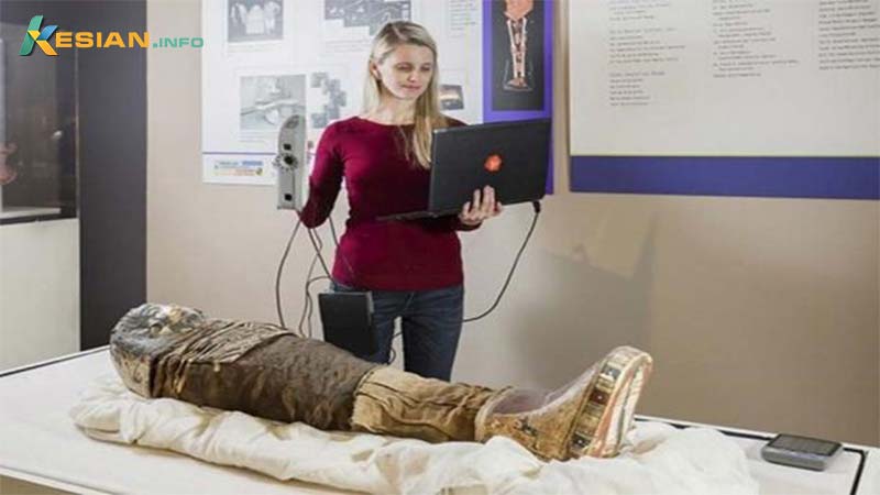
According to Live Science , scientists today call the child mummy “Sherit,” an ancient Egyptian name meaning “little one.” She lived in Egypt while the country was reigned over by the Romans and her burial style and the grave goods accompanying her body indicate she was part of a wealthy family . It is believed the girl died of dysentery or meningitis.
Julie Scott, executive director of the Rosicrucian Egyptian Museum in San Jose, California, where the child mummy is held, said why Sherit was chosen for the new project:
“For us, the value of this project is to bring this little girl’s story to life. She came to our museum in the 1930s, yet we knew very little about her. We wanted to find a way to learn more about who she was without damaging her mummy wrappings.”
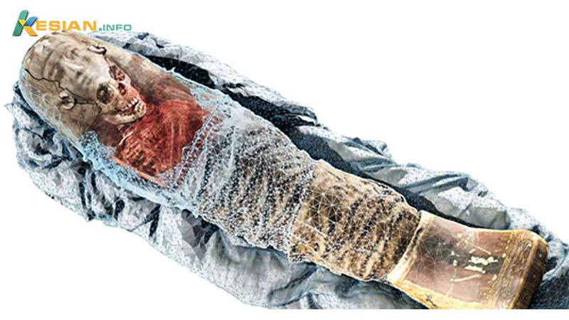
The combination of 3D scans with CT scans provide a whole new level of dimension to examining the mummy. The colorful details of the surface of the mummy provided by a handheld 3D scanner add depth to the images of a CT scanner – which complements the first with its ability to see beneath the wrappings.
Volume Graphics said “The result was, in this case, a mummy that is the most exact digital copy of the original to date, both within and without.”
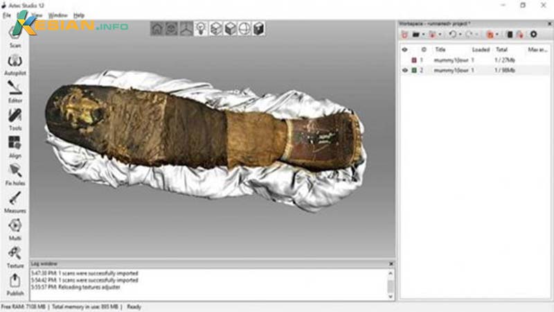
Live Science reports that the scans have been combined into a single 3D model using software developed by Volume Graphics. That software has been added to iPads in the Rosicrucian Egyptian Museum. Scott explained what the museum plans to do with this tool in a statement :
“Guests will be able to move an iPad over the mummy case, in order to see the associated scans. Our hope is that this new technology will help inspire guests to deeply relate to this little girl who lived so many years ago.”
Although the 3D and CT scan combo is currently in use for unravelling the story of the child mummy, the researchers suggest this tech will be useful for others working in archaeology, paleontology, biology, geology, and manufacturing as well.
Christof Reinhart, CEO of Volume Graphics, told Live Science it “allows for a more life-like, accurate representation of all kinds of objects and thus improves our understanding of these scanned objects.”


